In
the studies of plant pathology, it is define as a science that studies
the plant disease and the causes. This is included in the elements of
causal agents, mechanisms of infection and also the methods to control
those diseases. One of the steps that is need to be done studying plant
pathology is through the identification of plant disease on the host
plant. It involves in the determination of the environment conditions
and also the causal agents of the diseases. The identification is done
through the observation on the disease symptoms or signs of causal
agents on plant parts. Next is to know the techniques to isolate the
plant pathogens from diseased materials. This usually depends on the
growth, reproduction and ecology of the microorganism.
Other
than that, it is important to know the steps in Koch Postulates. The
association of a microorganism with a plant disease does not necessarily
proven that the microorganism is the causal agent. The identification
of causal agent should be confirmed by a related manually and if not,
the pathogenicity of the microorganism must be confirmed using the Koch
Postulates.
Objective
- To identify the disease of the plant through symptom observation.
- To differentiate the isolation techniques depending on the type of pathogen from diseased materials.
- To identify the disease of the plant through Koch Postulates.
Disease samples, Petri dish, slides, and culture media.
Methods
Activity 1: Identification of plant diseases through sign and symptoms
- Diseased samples were identified and observed
- The causal agents from diseased specimens were isolated
- The isolated causal agents were cultured in PDA plated and Na for further observation
- Pure culture of 2 fungal pathogens, Colletrichum truncatum and C.capsici , diseased chilli fruit, healthy chilli fruit, inoculation needle, plastics container, filter paper and PDA plates were given.
- Slides from both pure culture were prepared and observation was made under light microscope.
- The causal agent was isolated from the disease tissue into PDA plates by using aseptic technique. The plates at room temperature for observation in the next practical are labelled and incubated
- Aseptically, 1 agar block containing hyphae from each pure culture provided were cut and each species of pathogen on a sterilized chilli fruit were inoculate. The third chilli fruit are uninoculates as a control. The fruit in a moist tray were keep and covered with a pastics sheet. The fruit was incubate for 3 – 5 days and the symptom were observed.
- The isoaltion processes in (3) were repeat using fruit that demonstrated similar symptom as in (3) above (step 4).
Result and
Discussion
Activity 1:
Identification of plant disease through signs and symptoms observation
General necrosis:
Soft root
|
Most symptoms
are along the lines of watery and soft decay of the tissue.Often there is a
change in colour and in the case, the whole taproot can be decayed leaving just the
epidermis.
|
Vascular wilt
|
Avascular wilt
disease appears first as premature yellowing or other discoloration of the
leaves, while the stems and leaf petioles remain green. Plants may show
bunching of upper leaves and shortening of stem internodes, creating a
rosetting symptom.
|
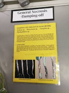
Damping-off
|
Affects seeds
and new seedlings, damping off usually refers to the rotting of stem and root
tissues at and below the soil surface. In most cases, infected plants will
germinate and come up fine, but within a few days they become water-soaked
and mushy, fall over at the base and die.
|
Blight
|
Symptoms include
sudden and severe yellowing, browning, spotting, withering, or dying of leaves, flowers, fruit, stems, or the entire plant.
|
Blast
|
Initial
symptoms appear as white to gray-green lesions or spots, with dark green
borders.Older lesions on the leaves are elliptical or spindle-shaped and
whitish to gray centers with red to brownish or necrotic border.
|
Dieback
|
Common symptom
or name of disease, especially of woody plants,
characterized by progressive death of twigs, branches, shoots, or roots,
starting at the tips. Staghead is a slow dieback of the upper
branches of a tree; the dead, leafless limbs superficially resemble a stag’s
head.
|
Local necrosis:
Scab
|
Leaves of affected plants may wither and drop
early. Plant diseases characterized by crustaceous lesions on fruits, tubers, leaves, or stems. The term is also used for the symptom of the disease.
|
Leave spot
|
Leaf spot
initially resembles drought or insect damage, and it can be difficult to tell
the difference.Leaf spot grows in random patterns on lawns, not in any
particular, recognizable shapes.
|
Anthracnose
|
Anthracnose
causes the wilting, withering, and dying of tissues. It commonly infects the
developing shoots and leaves.Symptoms include sunken spots or lesions (blight) of various colours in leaves, stems, fruits, or flowers, and some infections form cankers on twigs and branches.
|
Rust
|
Early on, look
for white, slightly raised spots on the undersides of leaves and on the
stems. After a short period of time, these spots become covered with
reddish-orange spore masses. Later, leaf postules may turn yellow-green
and eventually black. Severe infestations will deform and yellow leaves and
cause leaf drop.
|
Downy mildew
|
Older lesions
turn brown and appeared shrivelled. Mycelium of fungus forms mats and appears
as white, grayish white or tan colored patches on leaves, buds, stems or
young fruit. Fruiting bodies (cleistothecia) appear as small black or brown
specks on the mycelial mats. Infected leaves often appear chlorotic due to
decreased photosynthesis. Infected fruit and flowers are often aborted or
malformed. Early signs include small chlorotic spots or blistering on leaves
or flowers
|
Powdery mildew
|
Infected
plants display white powdery spots on the leaves and stems. The lower leaves
are the most affected, but the mildew can appear on any above-ground part
of the plant.
|
Canker
|
Symptoms
include round-to-irregular sunken, swollen, flattened, cracked, discoloured,
or dead areas on the stems (canes), twigs, limbs, or trunk. Cankers may
enlarge and girdle a twig or branch, killing the foliage beyond it.
|
Hypertrophy and hyperplasia:
Gall
|
Symptoms
include roundish rough-surfaced galls (woody tumourlike growths), several
centimetres or more in diameter, usually at or near the soil line, on a graft site or bud union,
or on roots and lower stems. The galls are at
first cream-coloured or greenish and later turn brown or black.
|
Smut
|
Smut is
characterized by fungal spores that accumulate in sootlike masses
called sori, which are formed within blisters in seeds, leaves, stems, flower parts, and bulbs. The sori usually break up into a black powder that is readily dispersed by the
wind. Many smut fungi enter embryos or seedling plants, develop systemically,
and appear externally only when the plants are near maturity. Other smuts are
localized, infecting actively growing tissues.
|
Witches broom
|
Can be easily
identified by the dense clusters of twigs or branches, which grow from a
central source resembling a broom. It is best seen on deciduous trees or
shrubs when they are not in leaf.
|
Hypoplasia:
Mosaic
|
Symptoms are
variable but commonly include irregular leaf
mottling (light and dark green or yellow patches or streaks). Leaves are
commonly stunted, curled, or puckered; veins may be lighter than normal or
banded with dark green or yellow. Plants are often dwarfed, with fruit and flowers fewer than usual, deformed, and
stunted.
|
Figure 3.0
After 4 days, the isolated causal agents from
diseased specimen (Chilli) have grown in the Petri Dishes respectively (Figure
3.0). However, the fungus that we cultured in the PDA is neither alike as ColletotrichumcapsicinorColletotrichumtruncatumbased on the
physical appearance of the pure culture (Figure 3.1) and (Figure 3.2)
Figure 3.1: Pure culture of Colletotrichumtruncatum
The slides (Figure 3.3) that we had made from our
cultured media show the mixture types of fungus instead of ColletotrichumcapsiciorColletotrichumtruncatum.
Therefore, this shows that the cultured media is contaminated with other type
of fungi.
Figure 3.3
Based on the slide of our cultured media, the
morphology of the fungus is not the same as the targeted fungus which are ColletotrichumcapsiciorColletotrichumtruncatumif compare to the
slide from the pure culture (Figure 3.4 &3.5). For example, the fungus does
not have the sickle-like shape conidia as in Colletotrichumcapsici.
Figure 3.5: Slide from pure culture of Colletotrichumtruncatum
Activity 3: Koch
Postulates
Pure culture
Figure 3.6
In Figure 3.6, this is the result of the Koch
Postulate process after 4 days. The healthy chillies that are wounded and inoculated
with the fungi disease now has shown the sign and symptoms of infection of Colletotrichum capsica and Colletotrichumtruncatumfungi.
Figures 3.8
The closer look of the chili that is inoculated with
Colletotrichumcapsici after 4 days(
figure 3.7 and 3.8). The infected chili started to show the infected symptom
which is sunken at infected part and growth of whitish mycelium within the
wound.
Figure 3.9
Figure 3.9 shows the closer look of chili that is
inoculated withColletotrichumtruncatum
for 4 days. The wounded part of the chili show little or no infected symptoms
of Colletotrichumtruncatum. This is
because the fungus pathogen, Colletotrichumtruncatumused
for inoculation is too little in the amount. Therefore, the infection process
of the pathogen require a longer period of time. Hence in Figure 3.9, the
wounded part of the chili did not appear the symptoms of infection.
Conclusion
In conclusion, disease in plant can identify though
observation on disease symptoms or signs of the presence of causal agents on
plant parts. In most diseases, the pathogens live or produce various kind of
structures on the surfaces of host. These structures include mycelia,
sclerotia, sporophores, fruiting bodies and spores which are called signs and
they are different symptoms which show visible responses on the infected part
of the host plant.


