Sunday, May 19, 2019

Introducing our group members for PLP3204


1. Toh Liang Su (199431)


2. Mohamad Asyraf Bin Ahmad Tajuddin (199300)


3. Jill Megan Chua Mei Chen (199392)


4. Mohd Nor Amiruh Shafarizan Bin Mohd Bukhari (199303)


5. Liu Lu (191333)


6. Amirul Ammar Bin Zainal Abidin (199370)

Monday, April 8, 2019

Lab Practical 8: Plant Virology

Introduction


In agriculture sectors, when it comes to the issue of virus control, it simply means to prevention is the only steps that should be done. The ultimate goal is to prevent virus spread simply just by either controlling the vectors or through the eradication of infected plants. In this also is an important step to prevent the farmers, agriculture industry  as well as the national and international trade. Viruses are sub-microscopic which is an infectious particles that multiply only inside living host cells. (Virology, n.d.) The basic shape of the viruses found in plants are elongate which is either in rigid rods or flexuous threads, cylindrical rods such as Bacillus-like and in isometric or polyhedral which is spherical shape. They are made out of two components which is a protein coat and also the nucleic acid center. They are also an obligate parasites which means they live in living tissues of plants before they reproduce themselves. They require a wound to gain entrance to a plant cell. In general, they severely affect the host both in quantity, quality and longevity. There are a few symptoms to extinguish the viruses which depends on a couple of factors such as:

1) Symptoms are important indicators of a plant virus disease.
2) Symptoms on a diagnostic indicator plants assists in characterizing the virus.
3) The ability to cause symptoms confirms the biological activity of the virus.

Objective

1. To study the diseases caused by virus.
2. To observe the morphology of virus.
3. To understand the steps in mechanical inoculation of virus on a host plant.

Materials

Figure 1 shows mortar and pestle

Figure 2 shows carborundum

Figure 3 shows the host plants which is chili plant

Figure 4 shows distilled water

Figure 5 shows diseased leaf


Mortar and pestle, carborundum, test host plant which is chili plant, distilled water and diseased leaf.

Procedure

Activity: Mechanical inoculation of virus on chili host plant.

  1. The virus inculum was prepared by crushing the diseased leaf by using the mortar and pestle until a uniform extract was obtained.
  2. 0.5ml to 1ml of distilled water was added to the extract.
  3. The host plant was dusted with carborundum abrasive on the leaf surface of the host which virus mix was inoculated.
  4. The carborundum caused injuries on the leaf surface resulting in the minute opening on the leaf surface. The carborundum was used only in a small quantity.
  5. The virus mix that has been prepared was applied on the test leaf surfaces of the host plant using a soft piece of cloth.
  6. Inoculation on the leaves was done by gently wiping the inoculum across the upper epidermis about 3-4 times to ensure that the leaves appear to be uniformly wetted and not damaged.
  7. The leaves that was inoculated with the virus was then washed using water. This was to remove excess leaf pieces and sap from the leaf surfaces. 


A. Virus Morphology

1. Chlorosis - An even change of colour which is yellow on the inside with the distinct spotting.


2. Necrosis - Cell death which appears as general necrosis or in the form of spots.


3. Mosaic - Characterized by light green, yellow or white areas intermingled with the normal green of the leaves or fruit. For example, mosaic of chili, tobacco and ground nuts.


4. Ringspot - Chlorotic or necrotic rings on the leaves and fruits.


5. Veinclearing - Chlorosis or necrotic rings on the leaves and fruits.


6. Veinbanding - severe chlorosis of the leaf veins. Example; Ageratum conzyzoides.


7. 'Penyakit Merah' of paddy.


Result and Discussion

The host plant of virus which is the chili plant was left within 24 hours in open air. After a day, observation and recording was done regarding the result of the infected chili host plant.


Chili host plant before inoculation


After 24 hours of inoculation
Left side - infected host plant
Right side - controlled host plant

Between the controlled chili host plant and the inoculated chili host plant, it was clear to state that the inoculation was experiment was successful as the infected host shows signs of inoculation. One of the obvious symptoms that can be seen is the curling up of leaves as compare to the control host. 


As shown in the picture above, the leaves that was inoculated with disease started to shows symptoms by curling up the leaves. The infection also doesn't seem to be spreading to other parts of the plants Other than that, the leaves started to turn yellow as shown in the picture but due to a day observation, the symptoms can't really be observe as the virus that was inoculated was a disease symptoms called mosaic.

Conclusion

In conclusion, virus can be seen through their morphology using the naked eyes such as chlorosis, necrosis, mosaic, ringspot, veinclearing and also 'penyakit merah' of paddy plant. This makes it easier for farmer to directly ifdentify the symptoms of viruses through the morphology of the symptoms. Other than that, we were able to understand the stages that is needed to be done to proceed the mechanical inoculation of virus on the chili host plants. It was clear that the virus require a wound to gain enterance to the plant cell. And also virus inoculation is fast thus the symptoms can already be seen within 24 hours after inoculation. The symptoms that can been seen through the inoculation was the curling of the leaves on the areas that was inoculated with viruses.

Reference

Plant Virology. (n.d.). Retrieved from FABI: https://fabinet.up.ac.za/research-groups/plant-virology

Virology, P. (n.d.). Plant Virology . Retrieved from Agricultural Research Council: http://www.arc.agric.za/arc-ppri/Pages/Plant%20Pathology%20and%20Microbiology/PLANT-VIROLOGY-.aspx


Monday, March 25, 2019

Lab Practical 7: Phytobacteriology

Introduction
Bacteria is one of the causal agent of disease in crops. Bacteria are one group of microorganisms that classified as prokaryote. The classification of bacteria depends on the shape, composition of cell wall and mobility. Bacteria can be divided into 3 main types according to shape, they are mainly coccus, bacillus and spiral. In addition, gram stain divides bacteria into 2 groups according to the composition of cell wall which are Gram positive (with peptidoglycan layer) and Gram negative (with peptidoglycan and an additional outer membrane). 
In general, bacteria reproduce by asexual method which is binary fission. It binary fission occurs every hour, after24 hours, 1 bacterium cell could reproduce approximately 16,800,000 new bacteria cells. In this lab practical season, the activities included: Isolation (direct and streak), Grain staining identification, KOH staining and Koch prostulate/ Diagnostic test.

Objectives
  1. To identify Gram positive and Gram negative staining under slides
  2. To observe the type of diseases caused by bacteria
  3. To perform the steps of isolation, KOH staining and Koch prostulate/ Diagnostic test
Materials
 Alcohol, chlorox, distilled water, diseased sample, potato, pen knife, culture media and inoculation needle.

Procedures
  1. Isolation (Direct)
  • The pen knife was wiped with 70% alcohol and the canker spots were cut out from the diseased sample by using the sterilized knife. The canker spots were dipped into 10% chlorox and distilled water before culture into the culture medium. The canker spots were cultured into NYGA and NA media.

    Figure 1
       2. Dilution streak
  • The same diseased tissues were crushed in distilled water for 10 minutes. Dilution streak was perform on the the provided Petri plates containing NYGA and NA medium.

 Figure 2
       3. KOH staining (Gram staining without dye)
  • One to two drop of 3% potassium hydroxide (KOH) was dropped on a slide. A loop of bacteria culture (E.coli & Bracillus sp.). The suspension of Gram negative bacteria has shown the sticky characteristic while Gram positive bacteria remained the same.

      Figure 3
      4. Diagnostic test using cut plant tissues
  • The skin of potato was peeled off and sliced into smaller pieces. The slice of potato was placed in sterilized Petri dishes. A "V" shape was cut on one slice and a drop of sterile water was put on it. This slice was labeled as control. Then, another "V" shape was cut in another slice of potato and it was inoculated with Erwinia caratovora on the "V". Both plates was incubated.

 Figure 4

Results and Discussions

Activity 1: Identification of Gram staning of bacteria

Figure 5.0: Gram negative (red)

Figure 5.1: Gram positive (Blue)

In figure 5.0, the Gram negative bacteria appear in red colour while the Gram positive bacteria retain in blue o purple colour. This is due to the Gram-positive bacteria take up the crystal violet stain used in the test, and then appear to be purple-coloured when seen through a microscope. This is because the thick peptidoglycan layer in the bacterial cell wall retains the stain after it is washed away from the rest of the sample, in the decolorization stage of the test. 

On the other hand, Gram-negative bacteria cannot retain the violet stain after the decolorization step; alcohol used in this stage degrades the outer membrane of gram-negative cells, making the cell wall more porous and incapable of retaining the crystal violet stain. Their peptidoglycan layer is much thinner and sandwiched between an inner cell membrane and a bacterial outer membrane, causing them to take up the counterstain (safranin or fuchsine) and appear red or pink.

Activity 2: Observation of disease symptoms
  1. Citrus canker, Xanthomonas axonopodis pv. citri
 
This bacteria infecting citrus leaves, branches and fruits. The symptoms including: sunken spot surrounded by yellow halo. However, the leaves are not wrinkled like those infected by scab.

     2. Bacteria wilt, Ralstonia solanacearum
The host of this bacteria included: chilli, brinjal, groundnut, okra, and tomato. Bacteria infecting the vascular tissues and causing symptoms of overall wilt in host plant.

     3. Soft rot of vegetable, Erwinia carotovora
This bacteria infecting various vegetables including: cabbage, brassica and carrot. Infected tissues turned watery, rot and produce bad odor.

     4. Stem rot of corn, Erwinia chrysanthemi pv. zeae
This bacteria infecting corn stem especially at the nod showing symptom of brown and water rotting.

     5. Black rot of crusifer, Xanthomonas campestris pv. campestris
This bacteria infecting various crucifer plants such as brassica and cabbage. The symptoms included dry rot starting at the leaf edge.

     6. Leaf blight of paddy, Xanthomonas oryzae pv. oryzae
The symptoms included: dry spots from the edge of leaves due to infection on hydathode openings.

     7. Leaf streak of paddy, Xanthomonas oryzae pv. oryzicola
 Symptoms: necrotic narrow stripes parallel to leaf veins.

     8. Bacteria spot of soy bean, Xanthomomas campestris pv. phaseoli
Symptoms: Small, water-soaked spots surrounded by a chlorotic halo appear on the leaves.

Activity 3: Isolation of bacteria
  1. Direct method
 
 Figure 6.0: Direct isolation of bacteria in NYGA media (left) and NA media (right)

Based on Figure 6.0, we can see the white mycelium growth of the disease causal agent in both NYGA medium (left) and NA medium (right). There are no significant differences between two culture medium as both of the medium produce similar result. Further process and isolation have to be  done to obtain the pure culture of the pathogen.

     2. Dilution streak method
 Figure 6.1: Dilution streak method of bacteria isolation in NYGA media (left) and NA media (right)

 Based on figure 6.1, we can see the growth of bacteria stain on both Petri plates (left) NYGA and (right) NA medium. Both medium produce similar results. However, there is contamination in NA media as we can see the growth of black colour spot surrounded by white mycelium. Procaution steps such as using sterilized tools, do the process faster to avoid contamination should be prioritized.

Activity 4: KOH staining
Figure 7.0: KOH staining on slides

Based on Figure 7.0, we can see that the Gram negative (bottom) appear to be viscous while the Gram positive (top) remain to be not viscous. This is because, KOH (Potassium hydroxide) dissolves the thin layer of peptidoglycan of the cell walls of gram negative bacteria, but does not affect gram positive cell walls. Disintergration of gram negative cell walls lyses the cell and release its contents, including the DNA. The DNA will make the solution very viscous and the solution will stick to the plastic loop when touched. Gram positive bacteria will not be affected by KOH, because they have thicker peptidoglycan layer in the cell wall. Thus, the cells will not be lysed, the DNA not released and no viscosity will be observed.

 Figure 7.1: Gram negative bacteria where the solution will be viscous and form a mucoid string.

Activity 5: Koch Prostulate
Figure 8.0: Infected potato with Erwinia sp (right) and control (left)

Based on figure 8.0, we can see that the infected potato slice show the rot symptoms of the tissues. The infected tissues turned watery, rot and produce bad odor. These symptoms is similar to soft rot caused by Erwinia caratovora. However, we can see that the control specimen (left) was contaminated. This maybe due to the use of contaminated sample or tools. Precaution steps such as using sterilized tools, do the process faster to avoid contamination should be prioritized. 

Conclusion
 In conclusion, plant diseases can be caused by bacterial infection. The examples of plant disease caused by bacterial including: Citrus canker, Xanthomonas axonopodis pv. citri, Bacteria wilt, Ralstonia solanacearum, Soft rot of vegetable, Erwinia carotovora, Stem rot of corn, Erwinia chrysanthemi pv. zeae, Black rot of crusifer, Xanthomonas campestris pv. campestris and etc. Bacterial isolation can be done by direct isolation or by dilution streak. In addition, the Gram staining is a common technique used to differentiate two large groups of bacteria based on their different cell wall constituents. The KOH staining is similar to gram staining which is to determine the gram negative and gram positive of bacteria.

References
  1. Wikipedia (online article). Gram positive bacteria. Retrieved from: https://en.wikipedia.org/wiki/Gram-positive_bacteria
  2. VetBact (online article). (2017). Potassium hydroxide test. Retrieved from: http://www.vetbact.org/index.php?displayextinfo=117


Monday, March 18, 2019

Lab Practical 6: Basidiomycota: Taxonomy and Pathological Importance


Introduction

One of the most considered developed phylum group is the Basidiomycota among the fungi consisting among the others of the mushroom, toadstools, puffballs and also stinkhorns. The rust and smut fungi are also included in the Basidiomycota although it is considered to be lower within the group. They are considered as plant pathogens that can cause millions of losses to annual crop production. The root disease pathogens that caused serious damages to many shade and other tree crops are also included. There are many mushroom that are edible and that contribute to the food industry. The Basidiomycota produces sexual spores called basidiospores which is usually 4 in number on a structure called the basidium and it may be formed fruiting body called the basidiocarp.

Objective
1.     To identify the type of specimens in under the phylum Basidiomycota under microscope.
2.     To observe the disease cycles of various basidiomycetes.
3.     To observe the type of disease caused by Basidiomycota on live specimens.

Result and Discussion

A.    Plant Disease Specimens
The specimen was examined and recorded the following symptoms:

1.     Horse hair blight, Marasmius equicrinus.


It is a minor problem found on stems of rubber, cocoa and etc. There is a presence of black hair-like rhizomorphs.


2.  White thread blight, Marasmiellus scandens.


Infected stems of plantation crops such as in cocoa and fruit trees crops. There is a white rhizomorph on the stems.


3.Panama disease or banana wilt, Fusarium oxyporum F.sp. Cubense.

The disease can attack young or old banana plant. The symptom can cause wilt on young banana plant. When the stem is cut opne, there is a black and brown colour in the stem.


4.1.     Sooty mould disease, Tripospermum sp. Capnodium sp. Limacinula sp.


The mould disease can cover up the mango leaf and in result prevent the tree to undergo photosynthesis and eventually wilt.


5. “Bintik daun antraknos Mangga”. Colletotricum gloesporioides.


Often found serious disease on mango trees. The younger plants can easily be affected compare to the older trees. Forms spots of brown colour on the fruits.


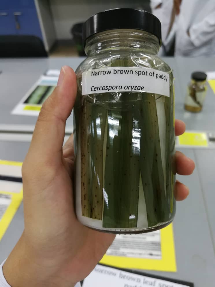
6.  Narrow brown spot of paddy, Cercospora oryzae.


The symptoms that is found is the presence of narrow brown spots on the paddy leaves.


7.    Citrus scab, Elsinoe fawcetti.

The presence of corky spot on the infected leaves which is looks like shredded and stunted growth.


8. Mouldy rot of rubber, Ceratocystis fimbriata.

Symptoms can be seen on the tapping area where there is spots of notched and the fungi can be seen as greyish white in colour.


9. Bird eye spot of rubber, Helminthosporium hevea.


The symptoms are falling of leaves, round spots near the veins on the leaves.


10. Back mildew, Meliola mangifera.


Presence of black mildew on the leaves with soot fungi on the leaves.


11. Leaf blight of maize/corn, Drechslera maydis.


Symptoms are leaf blight that is infected in maize plant where the blight colour is golden yellow to pale yellow.


12. Powdery mildew of rubber, Oidium heveae.


The leaves easily fall off and the leaves looks like it rolls and has a black tip.


13. “Keratan Basiodiocarps”


14. Brown spot of paddy, Drechslera oryzae.


Symptoms found are brown spots that appears on the leaves and also the panicles of the paddy plant.


15. Blast of paddy/ Rice blast, Pyricularia oryzae.


Consist of spots of molds on the leaves which is brown in colour.


16. False smut of paddy, Ustilaginoidea virens.


The fungi looks like a ball shaped with the spores that is yellow and green panicle. Eventually, the panicle becomes light and breaks when old.


17. Sigatoka disease of banana, Mycosphaerella musicola.


Symptoms are the leaves has lesion which is yellow in colour then turn brown with lines as it grows older.


18. Sheath blight of paddy, Rhizoxtonia solani.


The blight is normally forms spots on the leaves of the paddy plant. The colour to this blight id greyish green.


19. South American Leaf Blight, Microcylus ulei.


There is symptoms of black spots on the leaves and spreads to the edge of the leaves as it grows older.


20. Foot rot, Sclerotium rolsfiii.


The symptoms happens on ground such as in chili, tomato and other Solanaceae family. It can be seen on the bottom stem of the plant where rotting happens.
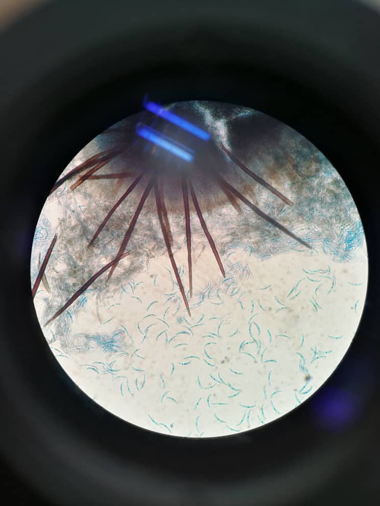

B. Slides


1. Phoma sp.


2. Botryodiplodia sp.


3. Saccharomyces cerevesiae


4. Collettotrichum gleosporiodes
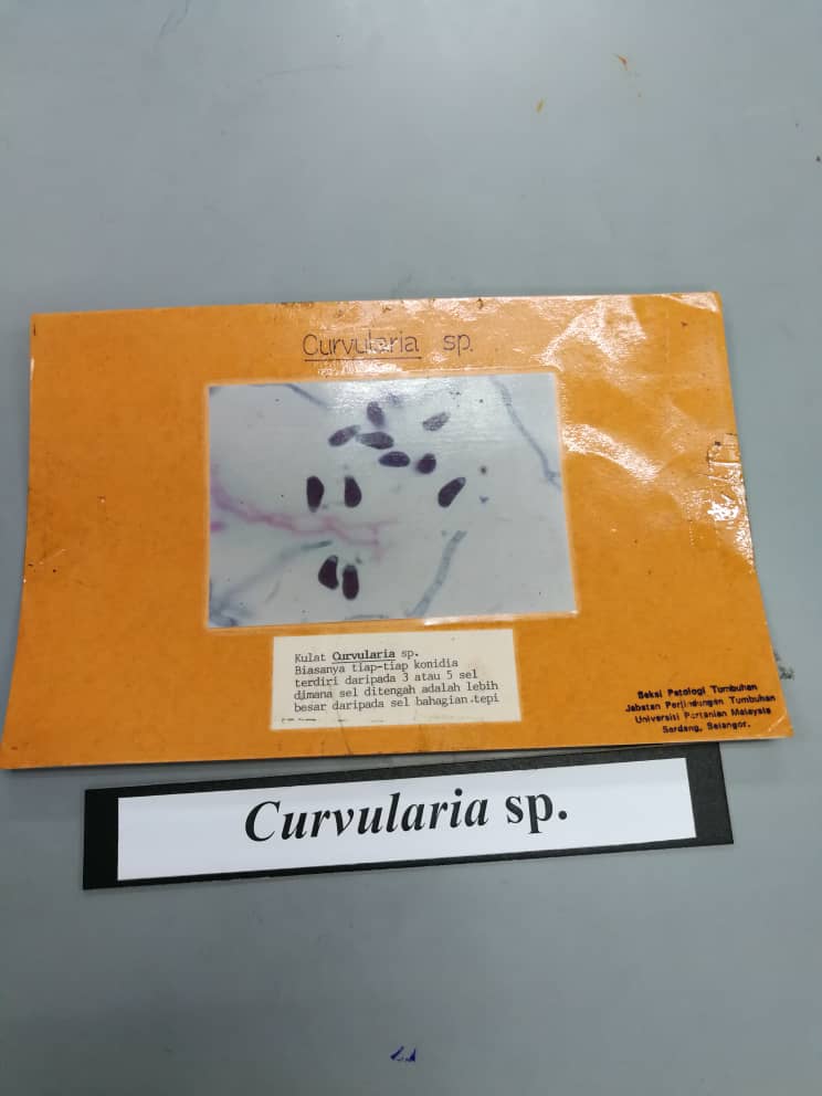

5. Curvularia sp.


6.  Colletotrichum capsici


7. Cercospora arachidicola


8. Stages in Puccinia graminis caused wheat rust.


Stage 0: Spermogonium and receptive hyphae on barberry leaf.


Stage 1: Aecium and eaciospores on barberry leaf.


Stage 2: Uredium and uredospores on wheat stem/leaves. Stage that shows the rust symptoms.


Stage 3: Telium and teliospores on wheat stems/leaves

The basidiospores are formed in stage 4 on wheat and this subsequently infects barberry.


9. Polyporales


10. Penicillium


11. Rhizoctonia sp.


12. Sclerotium sp.


Based on the observation regarding basidiomycota, we were able to identify the characteristics of each of the organisms. Some of the basidiomycota causes rust and also smut and other symptoms towards certain plant species. We were able to study the symptoms that causes the disease. Lastly, we were also able to know the stages in the Puccinia graminis which causes wheat rust. The wheat rust consist of 4 stages which each stages in different form the other. 


Conclusion

In conclusion, the diseases that is caused by the phylum of basidiomycota can be identified by looking through the symptoms that is visible on the plant. In the live samples, the disease called horse hair blight is caused by Marasmius scandens. The symptoms that can be seen through the live samples are the disease are black and hair-like rhizomorphs which is a minor probelm on stem of rubber, cocoa and so on. Other than that, basidiomycota which causes Puccinia graminis  was also observe in stages which is from stage 0 to stage 3.


Reference
Britannica, T. E. (n.d.). Basidiomycota. Retrieved from Britannica: https://www.britannica.com/science/Basidiomycota
Taylor, T. N. (2015). Learn more about Basidiomycota. Retrieved from ScienceDirect: https://www.sciencedirect.com/topics/agricultural-and-biological-sciences/basidiomycota