Bacteria is one of the causal agent of disease in crops. Bacteria are one group of microorganisms that classified as prokaryote. The classification of bacteria depends on the shape, composition of cell wall and mobility. Bacteria can be divided into 3 main types according to shape, they are mainly coccus, bacillus and spiral. In addition, gram stain divides bacteria into 2 groups according to the composition of cell wall which are Gram positive (with peptidoglycan layer) and Gram negative (with peptidoglycan and an additional outer membrane).
In general, bacteria reproduce by asexual method which is binary fission. It binary fission occurs every hour, after24 hours, 1 bacterium cell could reproduce approximately 16,800,000 new bacteria cells. In this lab practical season, the activities included: Isolation (direct and streak), Grain staining identification, KOH staining and Koch prostulate/ Diagnostic test.
Objectives
- To identify Gram positive and Gram negative staining under slides
- To observe the type of diseases caused by bacteria
- To perform the steps of isolation, KOH staining and Koch prostulate/ Diagnostic test

Procedures
- Isolation (Direct)
- The pen knife was wiped with 70% alcohol and the canker spots were cut out from the diseased sample by using the sterilized knife. The canker spots were dipped into 10% chlorox and distilled water before culture into the culture medium. The canker spots were cultured into NYGA and NA media.

Figure 1
2. Dilution streak- The same diseased tissues were crushed in distilled water for 10 minutes. Dilution streak was perform on the the provided Petri plates containing NYGA and NA medium.

Figure 2
3. KOH staining (Gram staining without dye)
- One to two drop of 3% potassium hydroxide (KOH) was dropped on a slide. A loop of bacteria culture (E.coli & Bracillus sp.). The suspension of Gram negative bacteria has shown the sticky characteristic while Gram positive bacteria remained the same.

Figure 3
4. Diagnostic test using cut plant tissues- The skin of potato was peeled off and sliced into smaller pieces. The slice of potato was placed in sterilized Petri dishes. A "V" shape was cut on one slice and a drop of sterile water was put on it. This slice was labeled as control. Then, another "V" shape was cut in another slice of potato and it was inoculated with Erwinia caratovora on the "V". Both plates was incubated.

Figure 4
Results and Discussions
Activity 1: Identification of Gram staning of bacteria

Figure 5.0: Gram negative (red)

Figure 5.1: Gram positive (Blue)
In figure 5.0, the Gram negative bacteria appear in red colour while the Gram positive bacteria retain in blue o purple colour. This is due to the Gram-positive bacteria take up the crystal violet stain used in the test, and then appear to be purple-coloured when seen through a microscope. This is because the thick peptidoglycan layer in the bacterial cell wall retains the stain after it is washed away from the rest of the sample, in the decolorization stage of the test.
On the other hand, Gram-negative bacteria
cannot retain the violet stain after the decolorization step; alcohol
used in this stage degrades the outer membrane of gram-negative cells,
making the cell wall more porous and incapable of retaining the crystal
violet stain. Their peptidoglycan layer is much thinner and sandwiched
between an inner cell membrane and a bacterial outer membrane, causing them to take up the counterstain (safranin or fuchsine) and appear red or pink.
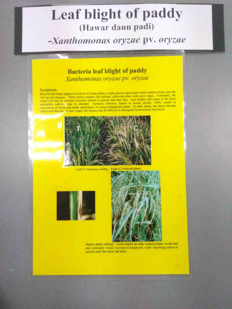
Activity 2: Observation of disease symptoms- Citrus canker, Xanthomonas axonopodis pv. citri

This bacteria infecting citrus leaves, branches and fruits. The symptoms including: sunken spot surrounded by yellow halo. However, the leaves are not wrinkled like those infected by scab.
2. Bacteria wilt, Ralstonia solanacearum

3. Soft rot of vegetable, Erwinia carotovora

4. Stem rot of corn, Erwinia chrysanthemi pv. zeae

5. Black rot of crusifer, Xanthomonas campestris pv. campestris

6. Leaf blight of paddy, Xanthomonas oryzae pv. oryzae
7. Leaf streak of paddy, Xanthomonas oryzae pv. oryzicola

8. Bacteria spot of soy bean, Xanthomomas campestris pv. phaseoli

Activity 3: Isolation of bacteria
- Direct method
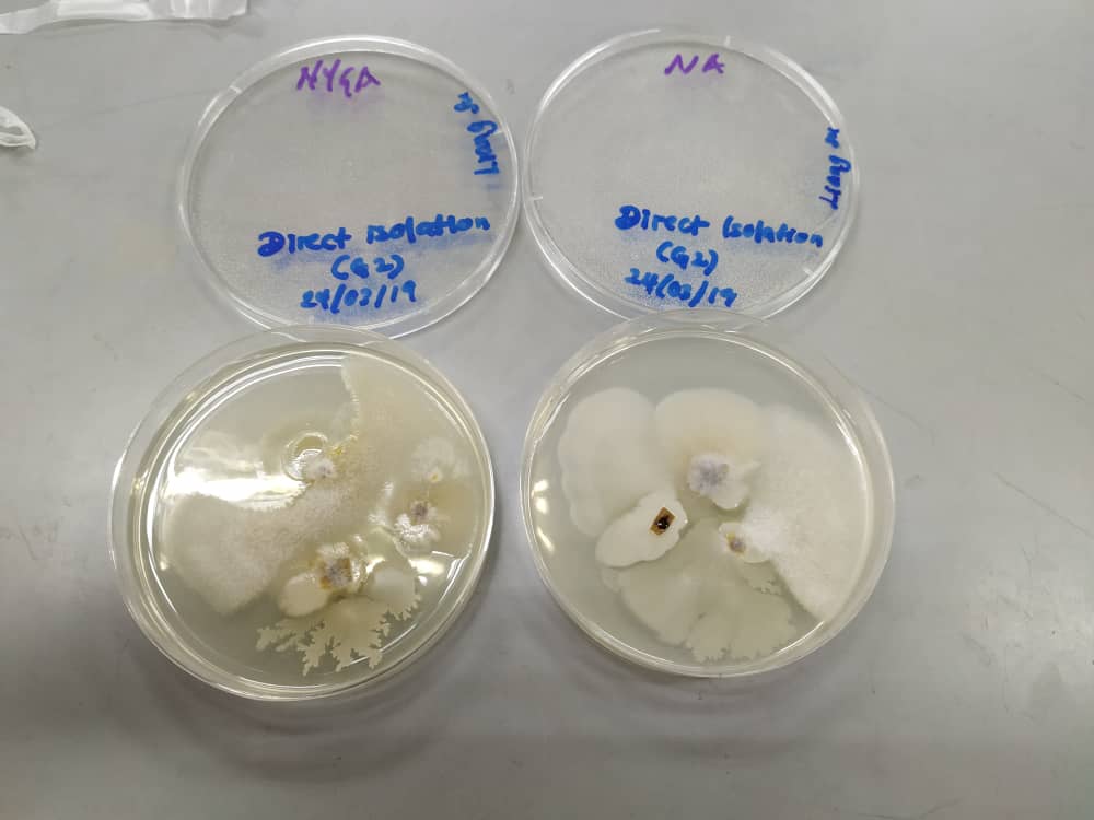
Figure 6.0: Direct isolation of bacteria in NYGA media (left) and NA media (right)
Based on Figure 6.0, we can see the white mycelium growth of the disease causal agent in both NYGA medium (left) and NA medium (right). There are no significant differences between two culture medium as both of the medium produce similar result. Further process and isolation have to be done to obtain the pure culture of the pathogen.
2. Dilution streak method

Figure 6.1: Dilution streak method of bacteria isolation in NYGA media (left) and NA media (right)
Based on figure 6.1, we can see the growth of bacteria stain on both Petri plates (left) NYGA and (right) NA medium. Both medium produce similar results. However, there is contamination in NA media as we can see the growth of black colour spot surrounded by white mycelium. Procaution steps such as using sterilized tools, do the process faster to avoid contamination should be prioritized.
Activity 4: KOH staining

Figure 7.0: KOH staining on slides
Based on Figure 7.0, we can see that the Gram negative (bottom) appear to be viscous while the Gram positive (top) remain to be not viscous. This is because, KOH (Potassium hydroxide) dissolves the thin layer of peptidoglycan of the cell walls of gram
negative bacteria, but does not affect gram positive cell walls.
Disintergration of gram negative cell walls lyses the cell and release
its contents, including the DNA. The DNA will make the solution very
viscous and the solution will stick to the plastic loop when touched.
Gram positive bacteria will not be affected by KOH, because they have
thicker peptidoglycan layer in the cell wall. Thus, the cells will not
be lysed, the DNA not released and no viscosity will be observed.

Figure 7.1: Gram negative bacteria where the solution will be viscous and form a mucoid string.

Figure 8.0: Infected potato with Erwinia sp (right) and control (left)
Based on figure 8.0, we can see that the infected potato slice show the rot symptoms of the tissues. The infected tissues turned watery, rot and produce bad odor. These symptoms is similar to soft rot caused by Erwinia caratovora. However, we can see that the control specimen (left) was contaminated. This maybe due to the use of contaminated sample or tools. Precaution steps such as using sterilized tools, do the process faster to avoid contamination should be prioritized.
Conclusion
In conclusion, plant diseases can be caused by bacterial infection. The examples of plant disease caused by bacterial including: Citrus canker, Xanthomonas axonopodis pv. citri, Bacteria wilt, Ralstonia solanacearum, Soft rot of vegetable, Erwinia carotovora, Stem rot of corn, Erwinia chrysanthemi pv. zeae, Black rot of crusifer, Xanthomonas campestris pv. campestris and etc. Bacterial isolation can be done by direct isolation or by dilution streak. In addition, the Gram staining is a common technique used to differentiate two large groups of bacteria based on their different cell wall constituents. The KOH staining is similar to gram staining which is to determine the gram negative and gram positive of bacteria.
References
- Wikipedia (online article). Gram positive bacteria. Retrieved from: https://en.wikipedia.org/wiki/Gram-positive_bacteria
- VetBact (online article). (2017). Potassium hydroxide test. Retrieved from: http://www.vetbact.org/index.php?displayextinfo=117
No comments:
Post a Comment